Ventral Slot Surgery Dog Recovery
By Dr. Joanne Fernandez-Lopez
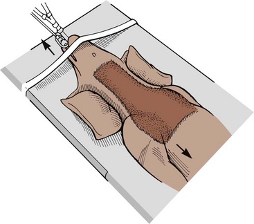
A triple ventral slot decompression surgery was performed. Not much is known about the factors that predict recovery in dogs with nonambulatory tetraparesis (NAT) secondary to cervical disk. Many dogs can have a complete recovery and return to normal function. Some dogs will be left with some hind limb weakness that never abates. There are some dogs that are so severely affected they do not require despite all our efforts. Many times these dogs can be managed in a cart. Hospitalization: These patients require around the clock care. A ventral slot is a surgery in which a hole centered on an intervertebral disc space is cut upward through the bone of two consecutive vertebral bodies to access the bottom of the spinal canal, enabling the removal of material compressing the spinal cord, such as extruded disc material, a blood clot, a cyst, an abscess, or a mass.

If your dog sustained a neck injury, you may be wondering about the dog neck surgery procedure and costs. In dogs, neck surgery is necessary for individuals with neurological deficits and conditions that could potentially cause permanent damage or when medical management (oral medications and strict rest recommended by a veterinarian) has proved to be unsuccessful. In dogs, there are two potential types of neck surgery options: Dorsal Cervical Hemilaminectomy and Ventral Slot Procedure. Veterinarian Dr. Fernandez goes over these dog neck surgery procedures and costs in the article below.
A Lesson in Anatomy
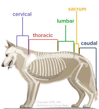
If we focus on the anatomy of the neck, the spinal cord lies within a chain of vertebrae bones. Each vertebrae bone has intervertebral discs. These are cushions or “shock absorbers” in between each vertebrae bone.
The discs may swell and rupture over time causing pressure on the spinal cord, severe pain and even inability to walk. Symptoms may vary depending on the amount of damage seen.
Minor spinal cord damage can lead to loss of coordination or “drunken walk.” More significant damage may show inability to move limbs voluntarily and loss of sensation in hind limbs.
The severe symptoms can carry a very poor prognosis for recovery depending on the duration that pain perception has been lost. If this occurs, it is an emergency and highly advice to visit the nearest veterinary emergency clinic as soon as possible.
A Matter of Breed
According to the American College of Veterinary Surgeons (ACVS), there are 2 types of breed groups in which one is more prone to develop symptoms than others.
They are grouped in chondrodystrophoid or non-chondrodystrophoid dogs. “Chondro” means cartilage and “dysplasia” means abnormal growth. Dogs that have abnormal development of the bone and cartilage are chondrodystrophic.
Chondrodystrophoid breed dogs include dachshunds, Pekingese, beagles and the Lhasa Apso. These dogs account for the vast majority of all disc ruptures, with Dachshunds accounting for 45 to 70 percent of all cases. The average onset of clinical signs is between 3 to 6 years of age.
Nonchondrodystrophoid dogs includeLabrador retrievers, German shepherd Dogs, and others. The condition presents between 5 and 12 years of age and the discs of the thoracolumbar (back region) account for 65% of all disc ruptures, while cervical (neck region) account for up to 18% of presenting cases
Signs of Trouble
Disc rupture presents with different degrees of pain; however, when nerve damage starts to develop and progress, it follows a predictable pattern:
Dogs with back or neck pain possibly refuse to walk around the room. They may develop a “drunken gait” or may get wobbly in the hind end. The hind feet will often cross as the pet steps.
There may be loss of hind limb motor function. Eventually the pet loses the ability to urinate and void (empty) their bladder completely. Loss of pain perception, which is a sign of severe cord injury that can carry a guarded to poor prognosis.
If your dog develops these symptoms see your vet immediately. Left untreated, disc rupture can lead to permanent loss of the ability to walk. Most dogs that reach this point will also lose control of their urinary bladder and are at risk for chronic urinary tract infections and urine scald. Additionally, without motor function, patients cannot turn themselves, and may develop bedsores and wounds.
At the Vet’s Office
Neurolocalization during physical examination will help the veterinarian choose diagnostics test and identify the potential surgery site. Classification of disc ruptures is generally grouped into large regions. The following groupings are described:
- Cervical vertebral or neck area 1–5 (C1–C5)
- Cervical vertebrae 6 through thoracic vertebrae 2 (C6–T2)
- Thoracic vertebrae 3 to lumbar vertebrae 3 (T3–L3)
- Lumbar vertebrae 4 through the sacrum (L4–S3).
Most primary care veterinarians may suggest initial health screening to determine any potential increase anesthetic risk or concurrent conditions. Diagnostic tests may include blood work ( complete blood count (CBC), serum chemistry) and a urinalysis.
Imaging techniques may include: X-rays of the neck and back, a myelogram, which is an x-ray series where a needle injects dye around the spinal cord to highlight any compression, a CT scan, magnetic resonance imaging (MRI) study, a spinal tap at the same time as the imaging. Your veterinary surgeon will determine the most appropriate tests, which may vary.
Dog Neck Treatment
Conservative treatment with cage rest, confinement, and pain medications is often only offered to patients that have recently begun their first episode and the neurologic deficits are mild. Further consultation with your veterinarian may result in a referral to a veterinary surgeon to fully explore your options.
Multiple diverse surgical procedures and approaches exist, varying on the veterinary surgeon and the location of the disc. Surgical decompression of the spine by removal of the bone over the spinal canal is nearly always recommended. Here I will briefly explain each procedure:
Dorsal Cervical Hemilaminectomy: This procedure is performed to gain access to the spinal cord through the dorso-lateral region of the spine or back. This approach is indicated when the lesion is located dorsal or lateral to the vertebral canal.
Ventral Slot Procedure: This procedure is performed to gain access to the ventral spinal cord in the neck region with ventral disc compression. With this procedure minimal manipulation of the spinal cord is necessary and recovery is generally rapid with few complications. Operation time is less compared to back surgery or hemilaminectomy.
Aftercare and Outcome
Most pets are discharged 3–7 days after surgery. Aftercare in the hospital is extremely important to improve outcome. The main goal is to provide supportive therapy, strict rest and pain management. Treated dogs are later sent home with oral medications, rest and then return for recheck and removal of skin sutures or staples.
Postoperative recovery following surgery may include: Bladder expression 3 to 4 times daily (if necessary), exercise restriction to “bed rest” for at least 4 weeks and physical rehabilitation for muscle strength and flexibility.
Life style changes may include weight loss, switching to a body harness instead of neck lead, and minimizing jumping off furniture.
Postoperative complications are possible and include:
- The myelogram could precipitate seizures in the first 24 hours after the procedure. This is why is important to keep patient in a 24/7 facility and provide anti-seizure medication if this complication occurs.
- Infection of the incision
- The possibility that the condition may recur. Many patients have another disc herniated later in life
- Continued wobbly walk or dragging hind toes when walking
Prognosis varies significantly with the degree of injury and the location of the injury. Most disc ruptures that present in dogs, still walking, have an excellent chance to return to walking. However, if the pet has lost the ability to sense pain in their legs before surgery is performed, they may never walk again.
Cost of Dog Neck Surgery
The average cost of cervical ventral slot or hemilaminectomy will vary based on standards of living as well as any additional costs incurred, including medications, laboratory tests, and postoperative supportive care.
When I used to work at a referral hospital, I have seen that the cost of diagnostics, surgical procedure, hospitalization and post-surgery care could range from $3,600 to $7,000 in severe cases.
References:
- Fossum T. W. et al. (2007) Small Animal Surgery. Chapter 38 Surgery of the Cervical Spine. Third Edition. Mosby ElSevier. Page 1402-1459
- ACVS. Intervertebral Disc Disease. Retrieved https://www.acvs.org/small-animal/intervertebral-disc-disease on July 18, 2017.
Photo Credits:
- Animal anatomical engraving from Handbuch der Anatomie der Tiere für Künstler’ – Hermann Dittrich, illustrator.
About the author
Dr. Joanne Fernandez-Lopez is an emergency veterinarian on staff in the Emergency and Critical Care Department at Florida veterinary Referral Center (FVRC).
Ventral Slot In Dogs

Originally from Puerto Rico, Dr. Joanne Fernandez-Lopez graduated from North Carolina State University – College of Veterinary Medicine in Raleigh, NC. Prior to joining FVRC, Dr. Fernandez-Lopez worked in small animal general practice and as a relief doctor in South East Florida. Her professional interests include dermatology, surgery, internal medicine, preventive medicine, reptile medicine and practice management.
In her free time, Dr. Fernandez-Lopez enjoys relaxing at the beach, paddle boarding, kayaking, and surfing. She has a small Tibetan spaniel mix named Carlitos.
Ventral Slot Surgery Dog Recovery Treatment


